HEMOGLOBINA
Informaçao geral
Textos

Informaçao especializada
Anemias
Catabolismo do heme


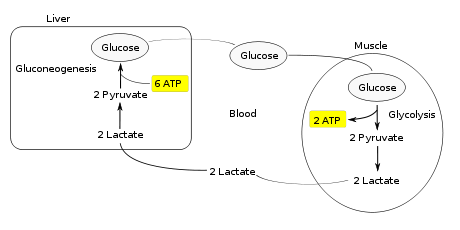
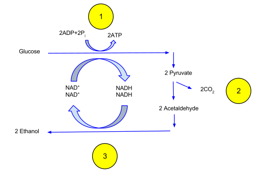
Ciclo de Rapoport-
Luebting
Genes da hemoglobina

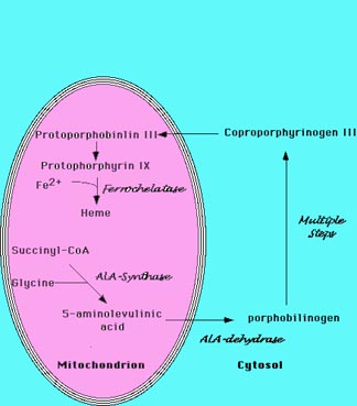
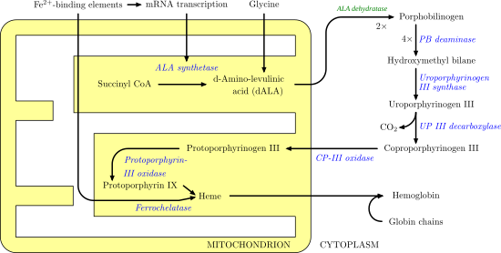
Heme


Texto e vídeo
Hemoglobina e
cooperatividade

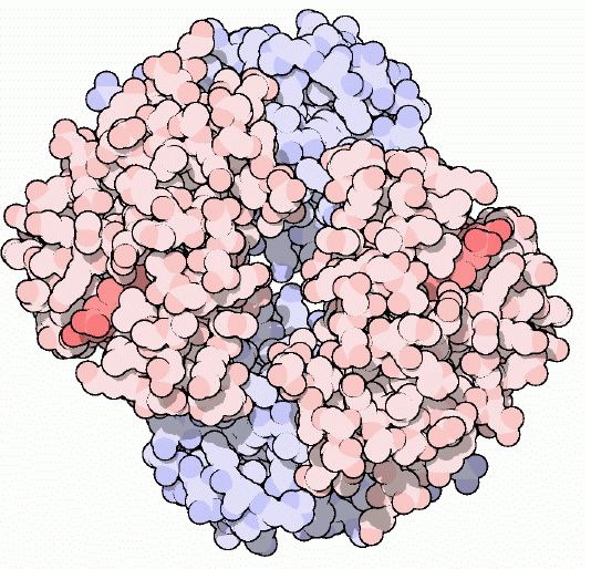
Hemoglobina
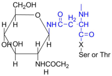
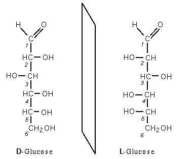
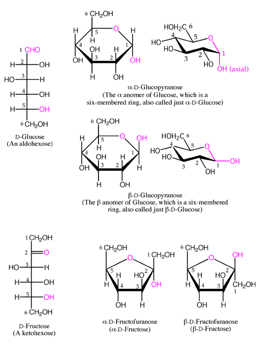
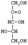
glicosilada
Hemoglobinopatias
Mioglobina


Porfirinas
Porfirias
Sintese do heme